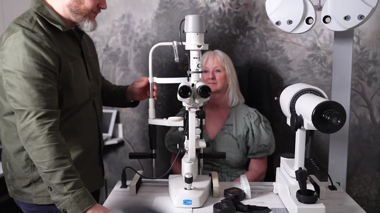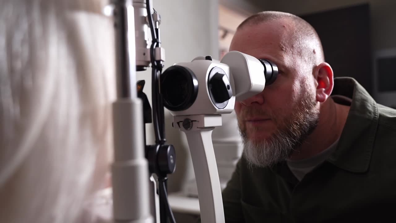
Optometry has changed more in the last fifteen years than in the fifty before it, and most of the change has been in what we can see rather than in what we can correct. A modern examination produces a record of the inside of your eye detailed enough to compare year on year — and the eye happens to be a uniquely informative place to look.
Why the eye is worth looking into
It is the one place where blood vessels and part of the central nervous system can be observed directly and non-invasively. The retina is nervous tissue; the vessels crossing it are the same vessels running everywhere else in your body. Changes in them are visible here before they are visible anywhere accessible.
What the technology adds
- Retinal photography — a wide, permanent image of the back of the eye, so this year can be compared with last
- OCT — a cross-section through the retinal layers, roughly analogous to an ultrasound scan of tissue a fraction of a millimetre thick
- Visual field testing — a map of your peripheral awareness, which is where several conditions show first
- Tonometry — the pressure inside the eye
- Anterior imaging — the tear film and the surface of the eye, which explains a great deal about comfort
What an eye examination can raise
Signs consistent with diabetes, raised blood pressure, and raised cholesterol can all appear at the back of the eye, and occasionally an eye test is the first place a systemic condition is noticed. We want to be careful about how that is phrased, because it is frequently overstated: an optometrist observes signs and refers. A diagnosis of a systemic condition comes from your GP with appropriate tests, not from us. What an eye test can detect goes through the specifics.
The part that is genuinely about eyes
The strongest case for imaging is not the systemic headline, it is the ordinary one: glaucoma, macular degeneration and diabetic changes in the eye are all conditions where early detection meaningfully changes what can be done, and all three are silent in their early stages. An OCT can show thinning at the optic nerve before a visual field test shows loss, and loss in the visual field is already permanent by the time it appears.
What it cannot do
It is not a health screen for your whole body, and it is not a substitute for blood tests, blood pressure checks or anything your GP does. Nor does a clear scan mean you do not have a condition it does not look for. It is one good window onto a small number of things, which is a useful and limited claim.
How we use it
Images are kept so that each examination can be compared with the last, because comparison is where the value is. If we see something that needs another opinion we refer, and we tell you what we saw and why. You can read about the technology at each practice on our eye examinations page.
The eye is a window, not an x-ray. It shows a few things very clearly and says nothing about the rest — which is worth being precise about.← Back to the Journal