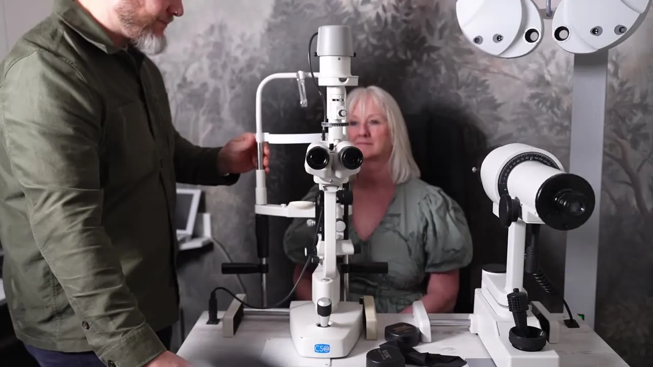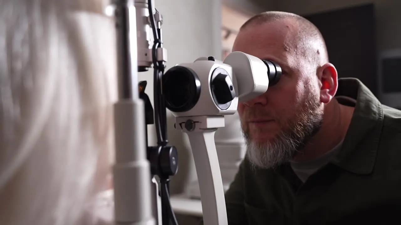
A standard eye test tells us a great deal. But some of the most important information about your eye health lies beneath the surface of the retina — in layers a conventional examination simply cannot see. That is where advanced imaging comes in.
A cross-section of your retina
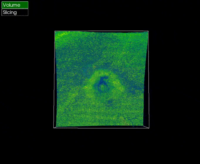
OCT — optical coherence tomography — is a quick, comfortable, non-invasive scan that captures a detailed cross-section of the layers beneath your retina. Think of it as the difference between looking at the surface of the ground and seeing a core sample of everything below it. It reveals detail that is invisible to the naked eye and to a standard sight test.
Why that detail matters
Several serious conditions — glaucoma, macular degeneration, diabetic changes — begin in these deeper layers, long before they affect your vision or become visible in a routine examination. Detecting them early gives you and your optometrist the earliest chance to monitor and manage them, and to protect your sight for the long term.
A picture you can build on
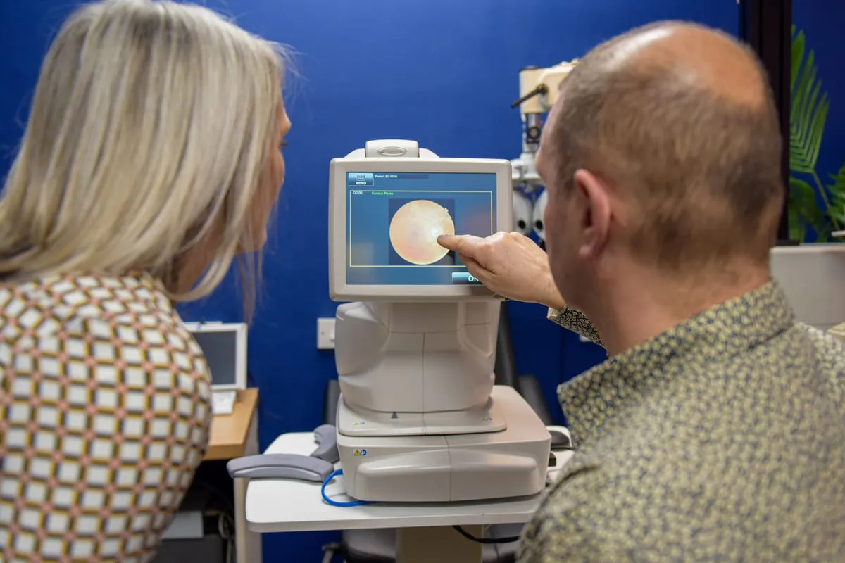
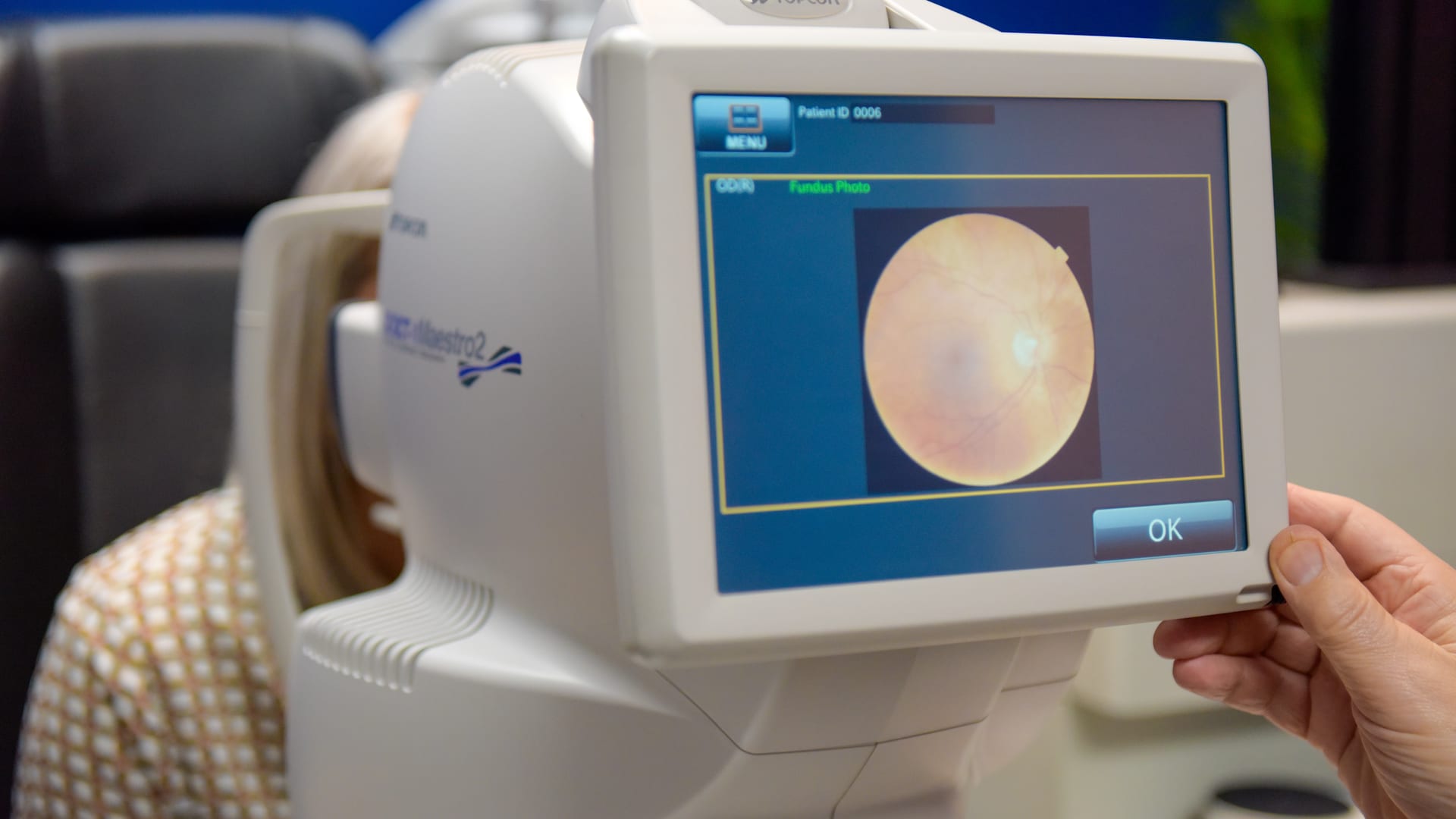
One scan is valuable. A series of scans over time is transformative. Because the imaging is so precise, we can compare year on year and spot subtle change with confidence — distinguishing what is stable from what needs attention. At our Urmston practice, pairing OCT with ultra-widefield Optomap photography, which captures a broad view of the back of the eye in a single image, gives us a remarkably complete picture of your eye health.
- Quick, painless and non-invasive
- In most cases, no dilating drops are needed
- Especially valuable from your forties onwards, or with a family history of eye disease
- Builds a year-on-year record that makes early change easy to see
Seeing deeper means acting earlier. That is the whole point of advanced imaging — to give you time, when time matters most.← Back to the Journal